- Retina Research Foundation
- About RRF
- Pilot Study Grants
- Grant Recipients 2025
- Samuel M. Wu, PhD
- Yingbin Fu, PhD
- Graeme Mardon, PhD
- Wei Li, PhD
- Yuqing Huo, MD, PhD
- Rui Chen, PhD
- Wenbo Zhang, PhD
- Curtis Brandt, PhD
- Lih Kuo, PhD
- Timothy Corson, PhD
- Jianhai Du, PhD
- Francesco Giorgianni, PhD
- James Monaghan, PhD
- Seongjin Seo, PhD
- Andrius Kazlauskas, PhD
- Erika D. Eggers, PhD
- Ann C. Morris, PhD
- Ming Zhang, MD, PhD
- Christine Sorenson, PhD
- Alex J. Smith, PhD
- Jeffrey M. Gross, PhD
- David M. Wu, MD, PhD
- Kinga Bujakowska, PhD
- Eric Weh, PhD
- Ching-Kang Jason Chen, PhD
- Jakub K. Famulski, PhD
- Thanh Hoang, PhD
- Georgia Zarkada, MD, PhD
- Eleftherios Paschalis Ilios, PhD
- Oleg Alekseev, MD, PhD
- Erika Tatiana Camacho, PhD
- Patricia R. Taylor, PhD
- Elizabeth Vargis, PhD
- Publications
- Grant Guidelines and Information
- Grant Application
- Grant Recipients 2025
- Research Programs
- Contact Us
- Giving
- RRF History
- Home
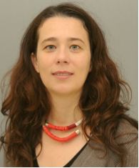
Kinga Bujakowska, PhD

Department of Ophthalmology
Ocular Genomics Institute
Massachusetts Eye and Ear
Harvard Medical School, Boston, Massachusetts
BASIC RESEARCH PROJECT
Modeling EYS associated retinitis pigmentosa in human iPSC derived retinal organoids
Research Interests
Retinitis pigmentosa (RP) is a group of inherited eye disorders that cause blindness to approximately 1.94 to 2.58 million people around the world. Mutations in EYS gene is one of the leading causes of RP. There is an urgent need to develop an appropriate human model for EYS to be able to study the function of this protein in photoreceptors, to understand the mechanism of the EYS-RP and test gene therapies. CRISPR-CAS9 edited human iPSC-derived ROs were used to model number of inherited retinal diseases, including RP, but EYS has never been studied in RO models before.
Plans for 2025
Mutations in EYS are one of the leading causes of RP 2–11. Despite the high prevalence of this disease, little is known about the role of EYS in the retina and the molecular mechanism of the EYS-associated disease. Dr. Bujakowska will use confocal microscopy and the high-resolutions expansion microscopy (Ex-M) to gain nanoscale insights into protein localization, particularly in the ciliary/periciliary structures of the photoreceptors in EYS RD retinal organoids and eys knock-out zebrafish. In light of a recent study suggesting that EYS is involved in the trafficking of the outer segment protein G protein–coupled receptor kinase 7 (GRK7)16, which plays a key role in photoresponse recovery, the lab will aim to further investigate alterations in the trafficking of other phototransduction proteins, particularly those critical to rod function, using our EYS-RD retinal organoids and eys knock-out zebrafish models.
Throughout 2025, Dr. Bujakowska will continue differentiating EYS-KO and wild-type retinal organoids and perform comprehensive immunohistochemical analysis using expansion microscopy (Ex-M) to explore nanoscale retinal anatomy and protein localization, focusing on ciliary structures. Her lab team will also investigate alterations in the trafficking of phototransduction proteins, particularly those important for rod function, in EYS-RD organoids and eys knock-out zebrafish, following recent findings on EYS involvement in GRK7 trafficking.
Specific Aims: Aim 1: To study the effects of EYS depletion on the development and maturation of retinal organoids. Aim 2: To study the involvement of EYS on trafficking of the phototransduction proteins.
Progress in 2024
In Aim 1, we collaborated with Aves Lab to develop a new anti-EYS antibody and successfully immunized hens, yielding antibodies with strong enzymatic activity as confirmed by ELISA. In Aim 2, CRISPR-based genome editing was used to create EYS knock-out iPSC lines, and retinal organoids are being generated to study the effects of EYS depletion on retinal development. EYS expression was detected in retinal organoids as early as day 70, and further characterization of EYS-deficient organoids will continue into 2025.
Mutations in the Eyes Shut Drosophila homolog (EYS) gene are the leading cause of RP in Asia and one of the five most mutated RP genes in the US and Europe. Despite the high prevalence of this disease, little is known about the role of EYS in the retina and the molecular mechanism of the EYS-associated disease. To address this knowledge gap, Dr. Bujakowska used CRISPR-Cas9 genome editing to establish human induced pluripotent stem cell (iPSC) lines harboring EYS frame shift mutations, thus yielding EYS knockout (EYS KO) cellular models and initiated the differentiation of these lines into retinal organoids (RO’s). In this funding period, the lab will conduct a comprehensive morphological examination of these retinal organoids using expansion microscopy (ExM) to gain nanoscale insights into retinal anatomy and protein localization, particularly concerning ciliary structures crucial for photoreceptor function in EYS deficient and wild type organoids.
For the next cycle, Dr. Bujakowska plans to continue EYS-KO and WT RO differentiation and will conduct a comprehensive immunohistochemical examination of these retinal organoids and human retina using the high-resolution expansion microscopy (Ex-M). Ex-M will provide nanoscale insights into retinal anatomy and protein localization, particularly of ciliary structures crucial for photoreceptor function.
Specific Aims:
Aim 1: To characterize EYS expression and localization in retinal organoids and human retina. Aim 2: To study the effects of EYS depletion on the development and maturation of retinal organoids.
Progress in 2023
In 2023, Dr. Bujakowska established a robust RO differentiation protocol from human iPSCs. She used the ROs to study expression and localization of EYS and different stages of development. Our principal objective was to elucidate the precise subcellular localization of EYS within retinal organoids and comparing it to the native placement in the human retina. Findings from this study were presented in a poster titled “EYS Expression in Human iPSC-Derived Retinal Organoids” at ARVO 2023 conference. The Bujakowska lab also used CRISPR-Cas9 genome editing to establish iPSC lines harboring bi-allelic EYS frame shift mutations, thus yielding EYS knockout (EYS KO) cellular models, and they are in the process of RO differentiation from these engineered iPSC lines.

